- Posted by MASTER VIKRANT ROHIN
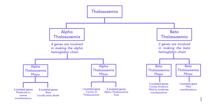
Thalassemia is an inherited blood disorder characterized by less oxygen-carrying protein (hemoglobin) and fewer red blood cells in the body than normal. it has two types Alpha and Beta.
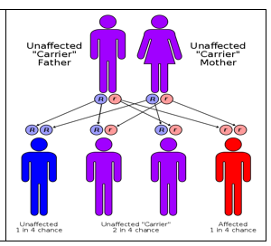
- Alpha and beta thalassemia are both conditions caused by genetic mutations.
- They both affect hemoglobin but in different ways.
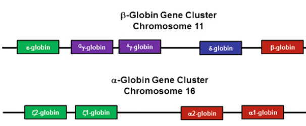
- Alpha thalassemia occurs when one or more of the alpha genes are deleted.
- Beta thalassemia occurs as a result of genetic changes to beta-globin genes.
- In both alpha and beta thalassemia symptoms may be absent or severe depending on which genes and how many are affected.
| TYPES : | TYPES : |
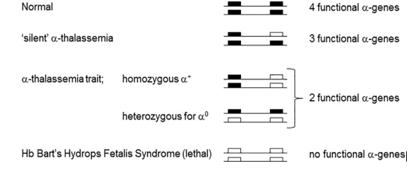
| There are two forms alpha thalassemia of depending on what genes are missing. Alpha + is when only one gene is missing from chromosome 16, while alpha 0 is when both genes are lacking from the same chromosome. | There are two types of beta thalassemia; one type is when only partial loss of function occurs. This is beta + thalassemia. The other type is the complete loss of function, which is beta 0 thalassemia. |
- There are different types of thalassemia, which can be divided into alpha and beta thalassaemias. Beta thalassaemia major is the most severe type. Other types include beta thalassemia intermedia,
THERE ARE 4 TYPES OF ALPHA THALASSEMIA:
- Alpha thalassemia silent carrier. One gene is missing or damaged, and the other 3 are normal. Blood tests are usually normal. …
- Alpha thalassemia carrier. Two genes are missing. …
- Hemoglobin H disease. Three genes are missing. …
- Alpha thalassemia major. All 4 genes are missing.
THERE ARE 2 TYPES OF BETA THALASSEMIA:
Beta thalassemia is classified into two types depending on the severity of symptoms:
- Thalassemia major (also known as transfusion-dependent thalassemia or Cooley’s anemia) and
- Thalassemia intermedia (which is a non-transfusion-dependent thalassemia). Of the two types, thalassemia major is more severe.
MAIN POINTS
| SYMPTOMS OF ALPHA THALASSEMIA: | SYMPTOMS: BETA THALASSEMIA: |
| In some cases there may be no symptoms and a person can be a carrier. This is often the situation where it is the alpha + form, and only one allele is affected. In other cases, where two alleles are missing, there are some symptoms such as smaller than normal red blood cells (microcytic anemia). In this case, there will be symptoms but they will not be too severe. If three of four alleles are missing then symptoms can be very bad and may include paleness, fatigue, swollen spleen, and jaundice. Such people may develop hemolytic anemia. If all four genes are affected a fetus will have Bart’s hydrops fetalis, and will not survive. | The symptoms vary depending on the extent of the damage to the genes. Individuals with few problems with beta globulin production are often silent carriers who show no symptoms. Some people are intermediate and may show some symptoms. Major beta thaleesemia (Cooley’s anemia), occurs when there are severe symptoms such as problems with the bone marrow, and severe anemia. Such patients can appear very pale; have jaundice, leg ulcers, enlarged spleens, and even gallstone formation. |
| DIAGNOSIS OF ALPHA THALASSEMIA: The diagnosis is based on the examination of a smear of blood that shows very small red blood cells. Fetal hemoglobin and hemoglobin A2 is usually normal in alpha thalassemia. Genetic testing can show deletions of alleles and electrophoresis of hemoglobin can show the condition as well. Fetal tissue can also be removed to test for the condition. | DIAGNOSIS: OF BETA THALASSEMIA Beta thalassemia is diagnosed by looking for hemolytic anemia by examining a red blood cell smear under the microscope. Testing of hemoglobin can be done and with severe beta-thalassemia, the hemoglobin would be low, less than 6 g/dL. There would be higher than average fetal hemoglobin and hemoglobin A2 in certain types of beta-thalassemia. Hemoglobin electrophoresis is used to diagnose beta-thalassemia. Hemolytic anemia is a blood condition that occurs when your red blood cells are destroyed faster than they can be replaced. |
TEST FOR KIDS
AMNIOCENTESIS. This is done from 15 weeks of pregnancy. A fine needle is passed through the mother’s tummy into the uterus to collect a small sample of the fluid surrounding the baby. The fluid contains some of the baby’s cells, which can be tested for sickle cell or thalassemia.
| CAUSES AND RISK FACTORS FOR ALPHA THALASSEMIA: The condition is inherited and is due to a genetic mutation of the alpha genes that are responsible for making alpha polypeptide chains. | CAUSES AND RISK FACTORS: BETA THALASSEMIA: This is inherited and is due to some type of genetic change (mutation) that has occurred in the beta-globin genes. |
| Region: This condition is more common in people who are of Southeast Asian, or African descent, or are from the Mediterranean regions of Cyprus and Greece. | Region: Beta thalassemia is more commonly found in people who are of Southeast Asian, African, or Mediterranean descent. |
| PREVENTION AND TREATMENT OF ALPHA THALASSEMIA: Genetic screening and counseling can help detect if there are genetic anomalies. Measuring hemoglobin levels in a couple may also help to show if they are carrying an alpha gene mutation. Sometimes a stem cell transplant, while the fetus is in utero, can cure them, or else the patient will need lifelong transfusions and potentially chelation therapy to remove excess iron from the blood. If the spleen becomes enlarged this may need to be surgically removed. |
| PREVENTION AND TREATMENT: BETA THALASSEMIA Genetic screening and hemoglobin testing can indicate if a person is carrying the mutation. Treatment may require blood transfusions, and chelation therapy to remove excess iron due to the transfusions, and in some cases, spleen removal is needed. |
- Chelation therapy involves weekly IV treatments of ethylenediaminetetraacetic acid (EDTA). Each treatment lasts about 30 minutes. In general, the medication seeks out and sticks to metals and minerals in the bloodstream, creating a compound that the body removes when urinating. केलेशन धातुओं को कमजोर लक्ष्य स्थलों से या उनके पास ले जाने में सक्षम बनाता है, और उनकी कार्सिनोजेनिक क्षमता को बाधित या सुगम बनाता है। विपरीत अर्थों में, धातु जटिल या मिश्रित जटिल गठन के माध्यम से लिगेंड सफाई करने में सक्षम हैं – बाद वाला बाइनरी कॉम्प्लेक्स के साथ बातचीत का परिणाम है।
DIFFERENCE BETWEEN ALPHA AND BETA-THALASSEMIA IN SHORT
- Definition: Alpha thalassemia is where there is a reduced formation of the alpha polypeptides. Beta thalassemia is where there is a reduced formation of the beta polypeptides.
- Symptoms: In alpha thalassemia, there can be no symptoms or in severe cases hemolytic anemia and an enlarged spleen, paleness, jaundice, and fatigue. In beta thalassemia, there can be no symptoms or in severe cases, there can be issues with the bone marrow as well as problems such as severe anemia, jaundice, gallstones, and enlarged spleens.
- Diagnosis: Alpha thalassemia is diagnosed by the presence of very small red blood cells in a red blood cell smear; genetic testing and electrophoresis of hemoglobin. Beta thalassemia is diagnosed by noting hemolytic anemia in a red blood cell smear; hemoglobin testing may indicate higher than average fetal hemoglobin, and hemoglobin A2 or lower than normal overall hemoglobin values.
- Causes: A mutation of one or more of the alpha genes on chromosome 16, causes alpha thalassemia, while a mutation of one or more of the beta-globin genes on chromosome 11 causes beta-thalassemia.
- Fetal Mortality: In the homozygous condition where all alpha genes are deleted the fetus develops Bart’s hydrops fetalis and dies in utero. In the homozygous condition of beta-globin genes, this is not fatal to the fetus in utero as there is some fetal hemoglobin that is formed.